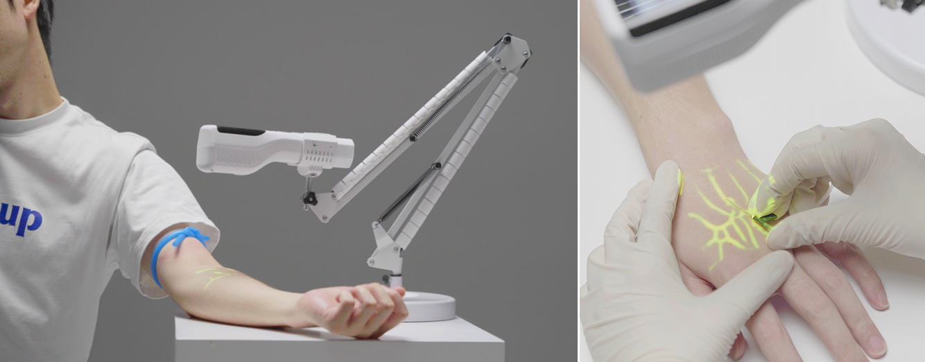
An infrared vein finder works by projecting near-infrared light (typically around 850 nm) onto the skin. Hemoglobin inside superficial veins absorbs this light far more strongly than the surrounding tissue, so the light reflected back to the device's camera creates a natural contrast pattern. A processor enhances this pattern in real time and a small projector displays the vein map directly onto the skin — exactly where the nurse needs it. The whole loop runs continuously, so the image moves with the patient's arm and updates with every heartbeat.
1. The Physics in Plain Language: Why Veins "Glow" in Near-Infrared
Every vein finder on the market exploits one biological fact: blood absorbs near-infrared (NIR) light differently from everything around it.
- Visible light (400–700 nm) reflects off the skin surface. That is why you can see tattoos and skin color — but not veins.
- Near-infrared light (roughly 700–1,000 nm) penetrates deeper into tissue. Within this window, hemoglobin — the iron-based protein inside red blood cells — absorbs NIR light strongly, with a useful absorption peak near 850 nm, while fat and connective tissue reflect most of it back.
Vein finder manufacturers, including ZD Medical, build their devices around this "biological window." The result: veins naturally behave like dark shadows against a bright background, without any contrast injection or dyes.
2. From Light to Image: The Four-Step Pipeline
A projection-based vein finder is not a camera and not a scanner. It is a closed loop that repeats dozens of times per second:
1. Illuminate. A ring of low-power NIR LEDs casts invisible near-infrared light onto the skin. Nothing is felt, nothing is injected.
2. Capture. An NIR-sensitive sensor records the light coming back. Veins appear dark (light absorbed by hemoglobin); surrounding tissue appears bright (light reflected).
3. Process. The onboard processor amplifies the contrast, filters out motion noise, and sharpens vein edges — including valves and bifurcation points, which are exactly the landmarks a nurse wants before a first stick.
4. Project. A visible-light projector paints the processed vein map back onto the skin, in real time and at 1:1 scale, so the green outline sits precisely over the real vein.
Because the loop is continuous, the projected image follows the patient's movement. Repositioning the arm does not "freeze" or invalidate the image — a practical advantage over still imaging methods.
3. What a Vein Finder Can See — and What It Cannot
Honest limits matter as much as capabilities, and knowing them prevents misuse:
Can see well:
- Superficial veins, typically in the upper ~8 mm of tissue — forearm, antecubital fossa, hand, wrist, and foot veins
- Veins invisible to the naked eye in pediatric, geriatric, oncology, and dark-skinned patients
- Vein valves, bifurcations, and "branching Y" points that help operators avoid risky puncture sites
Cannot do:
- It does not visualize deep veins or veins lying under significant edema or heavy adipose tissue — those belong to ultrasound
- It does not distinguish arteries from veins reliably, so it never replaces anatomical knowledge or palpation
- It does not confirm patency — a mapped vein can still be a fragile or previously-sclerosed vein; clinical judgment remains essential
This is why the light-vs-sound decision framework matters: a vein finder is the fast, first-line tool for routine and moderately difficult access; ultrasound is the escalation tool for deep or high-stakes access. They are colleagues, not competitors.
4. Handheld, Table Stand, or Trolley? One Principle, Three Configurations
The imaging principle is identical across all configurations; what changes is the workload and workflow of the department:
- Portable handheld units — the classic form factor: grab-and-go for ward rounds, home care, emergency response, and blood-draw teams. ZD Medical's portable projection vein finder is built around this use case, with the patented infrared projection technology at its core.
- Table stand models — for phlebotomy rooms, oncology day-care chairs, and pediatric clinics, where the puncture point is predictable and hands need to stay free. ZD Medical offers both a flexible table stand version and an enhanced table stand version.
- Mobile trolley models — for inpatient wards and multi-bed infusion areas, where one device serves many patients and needs to travel between beds. ZD Medical's vein finder with mobile trolley supports this workflow (support stands are also available separately, so teams can upgrade a handheld unit as needs grow).
Since 2012, ZD Medical has specialized in vein visualization products — this product line predates the company's ophthalmic imaging portfolio, and it is the area where our infrared projection technology has been refined longest.
5. Is Near-Infrared Safe Around Patients?
Yes. The NIR LEDs used in vein finders are non-ionizing, low-power light sources — the same portion of the spectrum used in household TV remotes, at intensities far below laser or phototherapy thresholds. No contact with skin is required, nothing is injected, and the light causes no heating sensation. This is why vein finders are routinely used on newborns, oncology patients receiving frequent infusions, and elderly patients with fragile skin — the populations where every avoided re-stick matters most, as we discussed in our article on first-stick difficulty in children and the elderly.
FAQ: What Clinicians Ask About Vein Finder Technology
Q: How deep can an infrared vein finder see?
A: Most projection vein finders reliably visualize superficial veins within roughly the top 8 mm of tissue; deeper vessels require ultrasound.
Q: Does a vein finder work on dark skin?
A: Yes — because it detects hemoglobin's near-infrared absorption rather than visible color, dark skin does not block the contrast, though very heavy pigmentation combined with edema can reduce clarity.
Q: Does a vein finder use radiation?
A: No. It uses low-power, non-ionizing near-infrared LED light — no radiation exposure, no injection, no skin contact.
Q: Can a vein finder replace ultrasound?
A: No — it is the faster first-line tool for superficial access, while ultrasound remains necessary for deep veins, arterial distinction, and guided cannulation of difficult targets.
Q: Why does the projected image include valve marks?
A: Valves and bifurcations create characteristic absorption shadows; showing them helps nurses choose a straight, patent segment and avoid puncturing at or below a valve.
Contact ZD Medical
Interested in the vein finder configurations mentioned above — handheld, table stand, or mobile trolley — or in requesting a demonstration for your department?
- Company: ZD Medical Inc. (Hangzhou, China)
- Email: sales@zd-med.com
- Tel: +86-25-51809558
- WhatsApp / Mobile: +86 187 9586 9515 (9:00–17:30, China time)
- Address: Room 301, 3rd Floor, Building 8, No. 19 Jugong Road, Xixing Street, Binjiang District, Hangzhou City, Zhejiang Province, China
- Website: www.zdmedimage.com


 compatible con la red ipv6
 español
 English
 العربية


